Publication on flexor tendon of the Toes

To date there has only been one published study describing the anatomy of the pulleys of the flexor tendons of the toes. The purpose of this article was to establish the existence of the pulleys to explain the pathophysiology of trigger toe in patients with stenosing tenosynovitis. The pulley system described had similarities to that seen in the fingers, but also some differences.
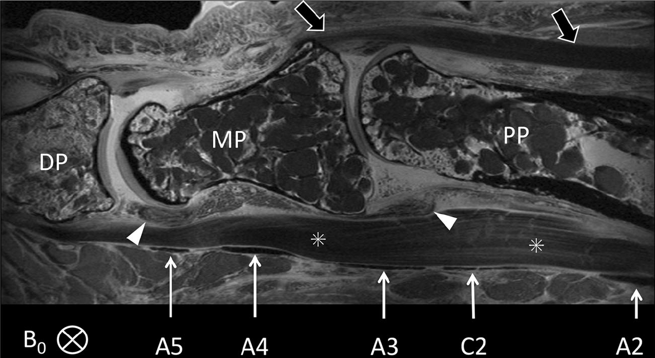
Sagittal fat-saturated SE scan (TR 3,500 ms/TE 15.7 ms) of the distal portion of the proximal phalanx (PP), the middle phalanx (MP) and the proximal portion of the distal phalanx (DP) of the 4th toe was obtained. The annular (A2, A3, A4, A5) and cruciate (C2) pulleys are seen in cross section as thin hypointense bands (arrows) superficial to the flexor tendons (?). The A2 pulley is only partially shown. The A2 and A4 pulleys are thicker than the cruciate (C2), A3 and A5 pulleys. The relations of the pulleys to the phalanges, interphalangeal joints and plantar plates (arrowheads) can readily be seen. B0 was perpendicular to the longitudinal axis of the toe and in the ML direction. The distal portion of the extensor tendon and its insertion on the base of the MP are also demonstrated (open arrows)