Publication on Novel MRI of the Meniscus

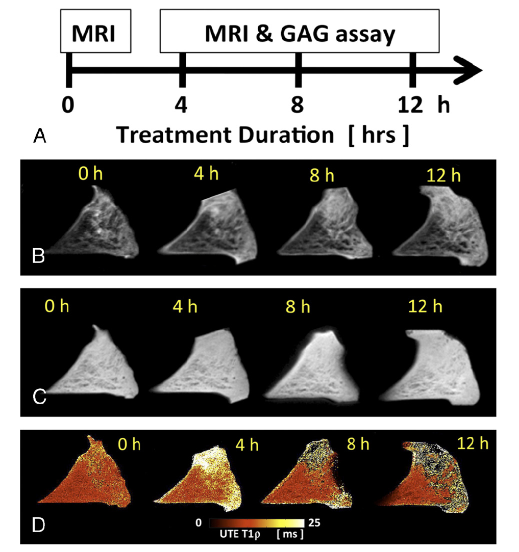
As novel and quantitative MR techniques become available, their clinical applications need to be validated. The aim of the study was to determine whether the novel quantitative ultrashort echo time (UTE) T1rho MR measurements are sensitive to proteoglycan degradation in human menisci induced by trypsin digestion. We used dissected meniscus samples. MRI was performed before and after 4, 8, and 12 hours of trypsin solution immersion, inducing proteoglycan loss. MR images showed progressive tissue swelling, fiber disorganization, and increase in signal intensity after GAG depletion. The UTE-T1ρ values tended to increase with time after trypsin treatment (P = 0.06). Cumulative GAG loss into the bath showed a trend of increased values for trypsin-treated samples (P = 0.1). We concluded that UTE T1rho measurements can non-invasively detect and quantify severity of meniscal degeneration.

A, Study design showing enzymatic treatment times. B, Meniscal morphologic and signal changes on PD-weighted imaging, after progressive enzymatic GAG digestion. The image at t = 0 hour (before enzymatic digestion) depicts the detailed collagen fiber network as well as the normal triangular shape of the meniscal horn. While the trypsin digestion time progresses (from t = 4–12 hours), progressive shape and contour changes are observed (secondary to swelling), as well as fiber disorganization and increase in signal intensity (related to GAG depletion). C, UTE-T1ρ images profile similar morphologic changes with increasing digestion times but clearly have increased signal as compared with the PD-weighted images. D, UTE-T1ρ color maps show altered quantitative values corresponding to the morphologic changes.


