Publication on TFCC of the Wrist
 The triangular fibrocartilage complex (TFCC) is the main biomechanical stabilizer of the distal radioulnar joint of the wrist. In this study, we evaluated pathology of the TFCC tissues using high-resolution morphologic magnetic resonance (MR) imaging, and compare
with quantitative MR and biomechanical properties in 5 cadaveric wrists.
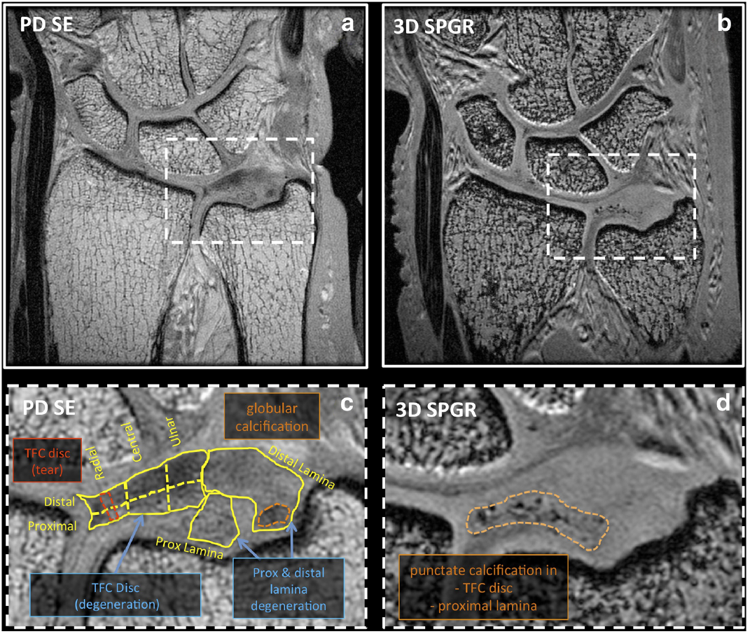
On morphologic PD SE images, TFC disc pathology
included degeneration and tears, while that of the laminae
included degeneration, degeneration with superimposed tear,
mucinous transformation, and globular calcification. Punctate
calcifications were highly visible on 3D SPGR images and
found only in pathologic regions. Disc pathology occurred
more frequently in proximal regions of the disc than distal
regions.
Quantitative MR values were lowest in normal samples,
and generally higher in pathologic regions. Biomechanical testing demonstrated an inverse relationship, with indentation modulus being high in normal regions with low MR values. These results show technical feasibility of morphologic MR, quantitativeMR, and biomechanical techniques to characterize pathology of the TFCC. QuantitativeMRI may
be a suitable surrogate marker of soft tissue mechanical properties,
and a useful adjunct to conventional morphologic MR techniques.