Morphologic MRI may not detect subtle and slow changes in tissue that may occur with degeneration, remodeling or healing.
Morphologic
Knee
Clinical Challenge
Technical Approach
Use novel sequences such as Ultrashort Time-to-Echo (UTE) MRI and high spatial resolution afforded by latest , and the newest morphologic techniques tailored for small tissues.
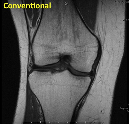
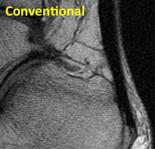
Knee Conventional
This conventional MRI of a knee shows articular cartilage and menisci with medium to low signal intensity.
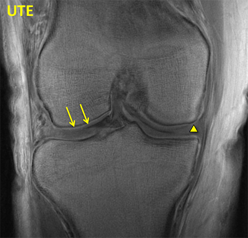
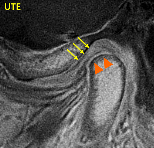
Knee UTE
UTE MRI reveals deep and calcified layers of articular cartilage (arrows) and menisci (triangle) with high signal intensity.
Spine
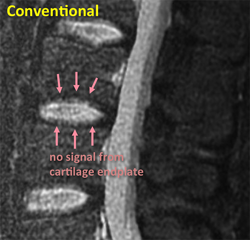
Spine Conventional
This conventional MRI of a spine shows cartilage endplates with low signal intensity, making it difficult to evaluate them.
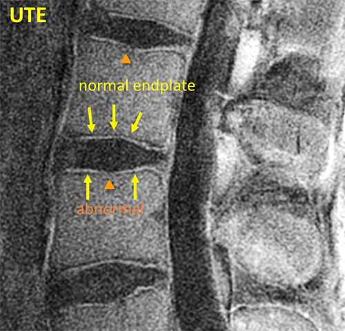
Spine UTE
UTE MRI reveals the structure of cartilage endplates with high contrast. Both normal and abnormal structures could be discerned here.
Wrist
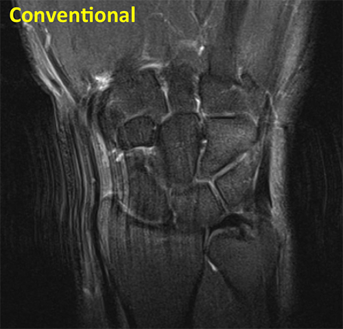
Wrist Conventional
Conventional coronal fluid sensitive sequence of the wrist shows intermediate signal of articular cartilage, and low signal in the triangular fibrocartilage (TFC).
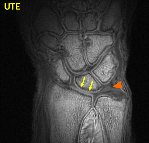
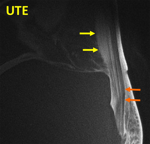
Wrist UTE
UTE sequence of the wrist shows bright signal in the deep and calcified layer of cartilage (arrow) and in the triangular fibrocartilage (arrowhead) (TFC).
TMJ
TMJ Conventional
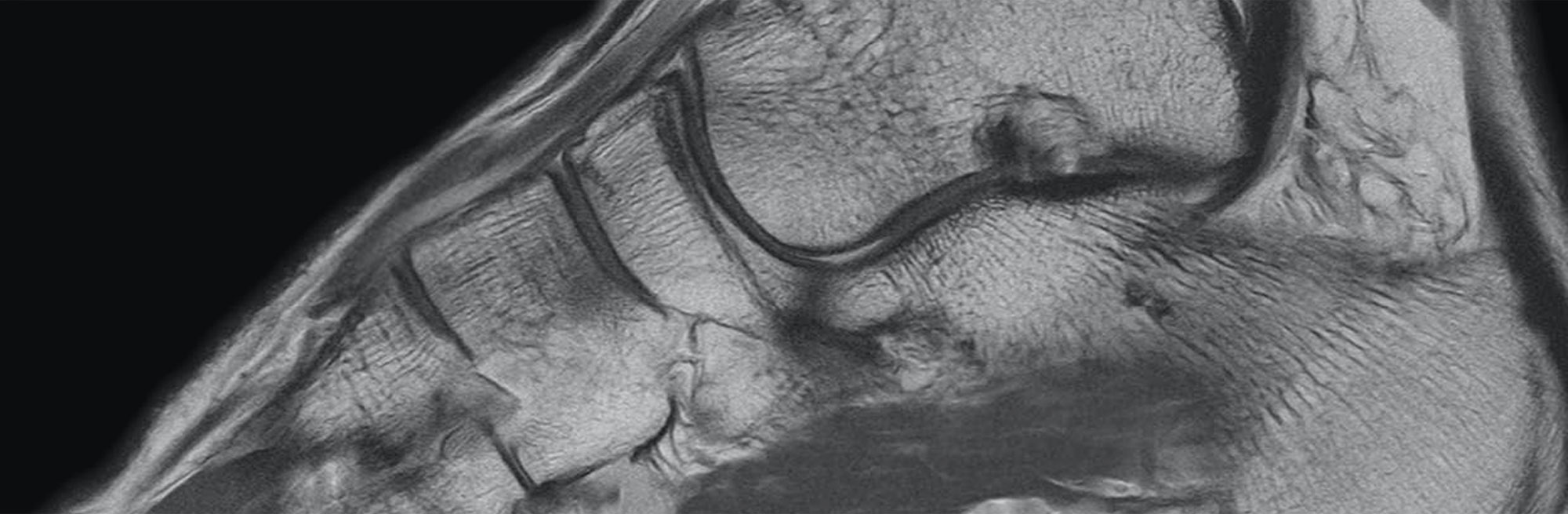
Conventional sagittal fluid sensitive sequence of the TMJ shows the low signal intensity disc and the mandibular condyle.
TMJ UTE
UTE sequence of the TMJ shows bright signal in the disc (arrows) and in the fibrocartilage covering the mandibular condyle (arrowhead).
Tendon
Tendon Conventional
Conventional sagittal MR image of the ankle shows the low signal intensity Achilles tendon.
Tendon UTE
UTE sequence of the ankle shows normal fasciculation (white arrow) in the tendon and a region of tendinosis (orange arrow) that could not be seen on the standard MRI.