Morphologic MRI may not detect subtle and slow changes in tissue that may occur with degeneration, remodeling or healing.
Quantitative
Knee
Clinical Challenge
Technical Approach
Use quantitative methods to determine tissue's intrinsic MR properties, such as T1, T2, and T1rho values. Our research focused on correlating MR properties with biomechanical and biochemical properties of MSK tissues.
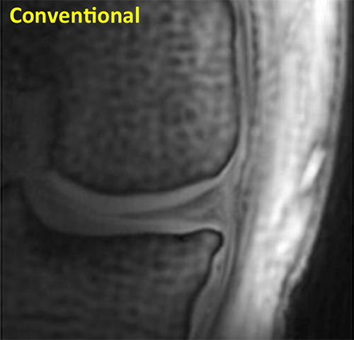
Knee Conventional
This conventional MRI of a knee shows articular cartilage with uniform signal intensity.
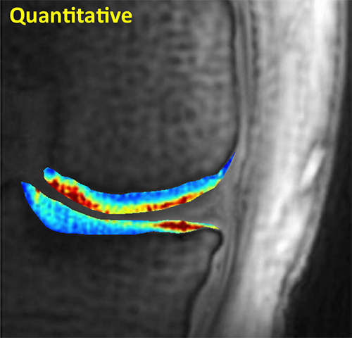
Knee Quantitative
Quantitative MR reveals regional variations in T2 values of articular cartilage with high sensitivity.
Spine
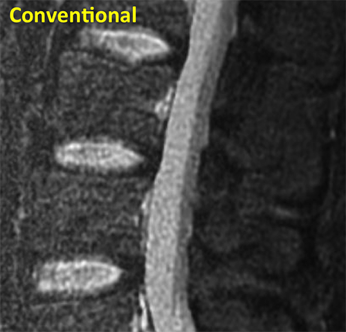
Spine Conventional
This conventional MRI of a spine shows three intervertebral discs with similar signal intensities.
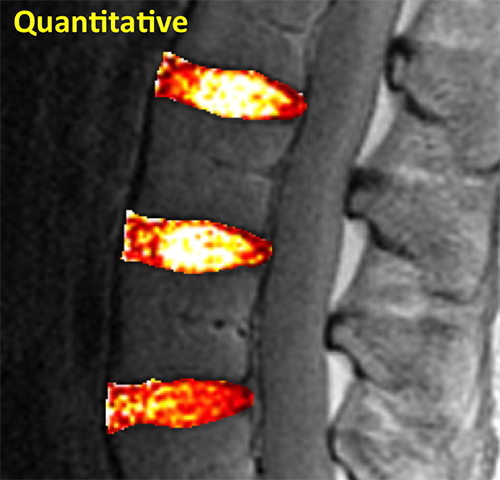
Spine Quantitative
Quantitative MRI reveals subtle differences in T2 values of intervertebral discs, along with internal variations.
Wrist
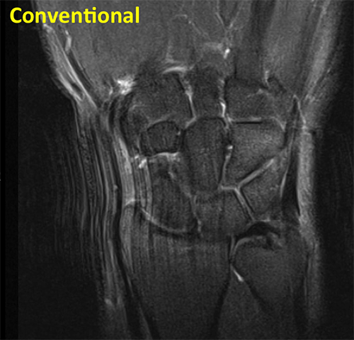
Wrist Conventional
Conventional coronal fluid sensitive sequence of the wrist shows intermediate signal of articular cartilage, and low signal in the triangular fibrocartilage (TFC).
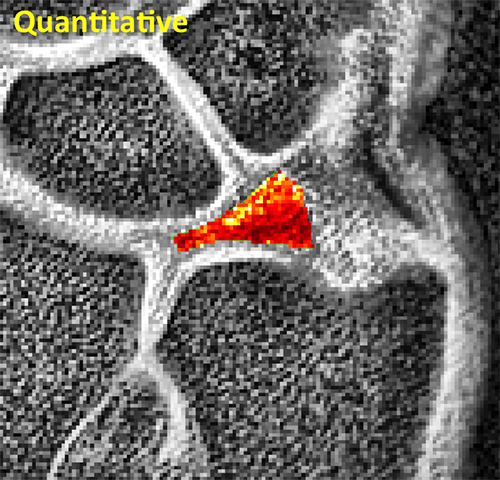
Wrist Quantitative
Quantitative MRI reveals shorter T2* values in the proximal portion compared to the distal portion of the triangular fibrocartilage complex (TFCC)
TMJ
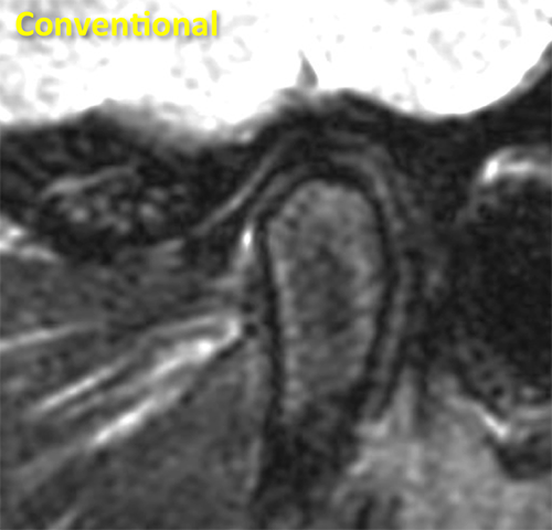
TMJ Conventional
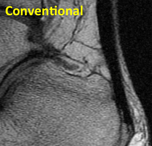
Conventional sagittal fluid sensitive sequence of the TMJ shows the low signal intensity disc and the mandibular condyle.
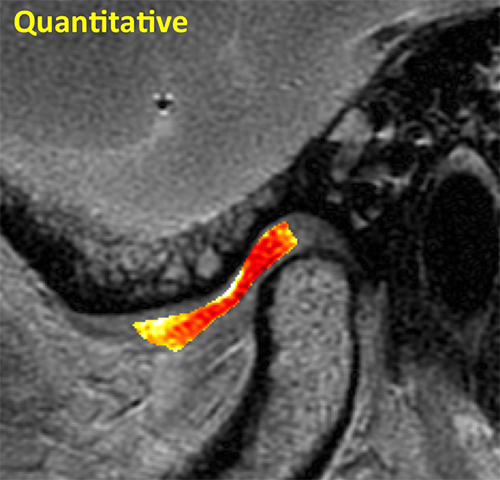
TMJ Quantitative
Quantitative MRI reveals subtle differences in T2 values of the TMJ disc that reflect early changes in its collagen structure.
Tendon
Tendon Conventional
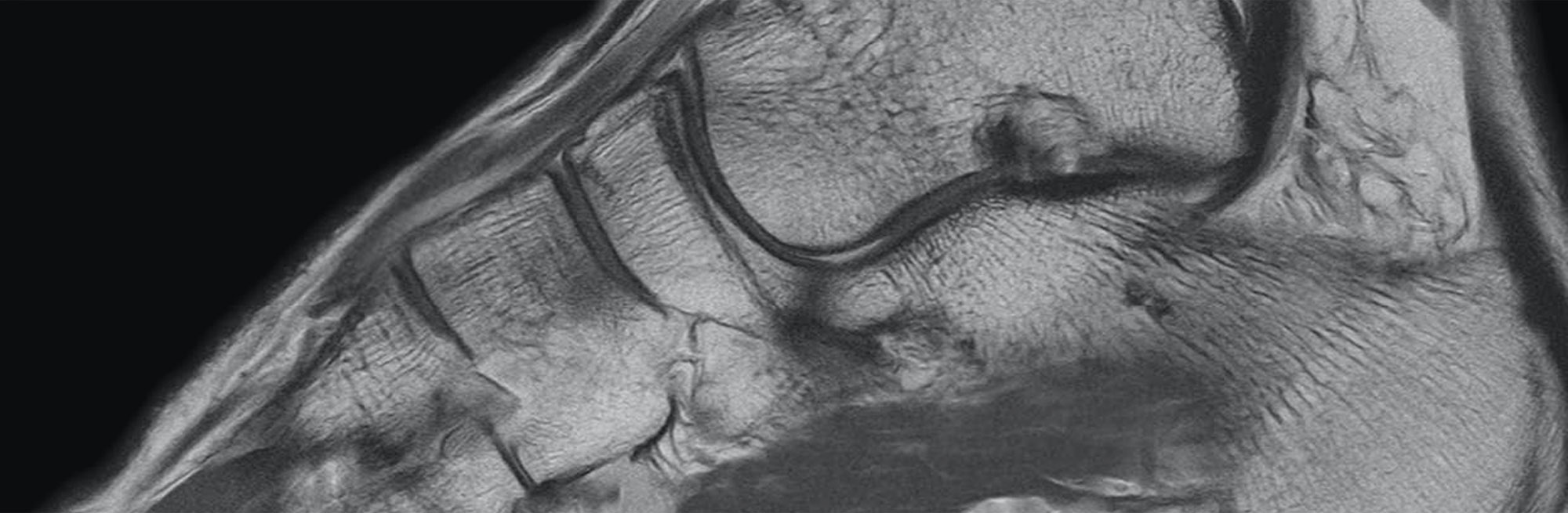
Conventional sagittal MR image of the ankle shows the low signal intensity Achilles tendon.
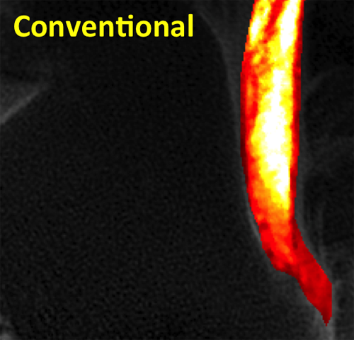
Tendon Quantitative
Quantitative MRI reveals marked changes in T2 values with bright signal centrally representing collagen fiber disorganization.